Diagnosis and Imaging (X-Rays and Scans)

Breast imaging is essential throughout the patient pathway:  

  • To help with diagnosis  
  • To ‘stage’ the disease  
  • To see if the cancer has spread and where in the body 
  • To assess response to treatment  
  • In follow-up to make sure the cancer has not come back (recurrence) 

The Scottish consensus on breast cancer imaging has been adapted from the UK guidance written by the Royal College of Radiologists (RCR). It provides advice on which imaging may be considered, including X- rays and mammograms, ultrasound scans (USS), CT (computerised tomography), MRI (magnetic resonance imaging) scans, PET scans (positron emission), and other tests.  The guidance explains which test to use and when, to give the best information to your healthcare team.  

The RCR guidance also includes advice for male breast cancer and pregnant or breastfeeding women where X-ray exposure is to be avoided.
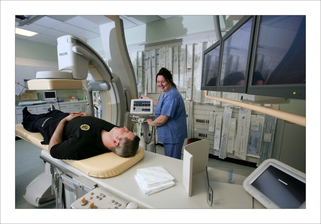
Patient having X-ray
If you are suspected to have breast cancer there are usually three types of tests to help diagnosis

– An appointment and examination with a doctor or nurse 
Mammogram (breast X-rays) 
Biopsy: usually sample taken by needle  

If a mammogram has been done within 6 months and is normal, or the examination does not find signs of cancer, an ultrasound scan of the breast may be helpful. Ultrasound is also preferred in pregnant or lactating women to avoid X-ray exposure. 
 
If cancer is found, another ultrasound may be used to check if the cancer has spread into the glands (lymph nodes) in the armpit (axilla) which is the usual first point of spread.  If it looks like lymph nodes are involved a biopsy may be taken from there too. 

Sometimes small ‘markers’ or ‘clips’ will be inserted into the areas of biopsy.  These markers are small and will not cause harm or discomfort.  These markers can show up on scans to help review these areas. 
 
Magnetic Resonance Imaging (MRI) is sometimes used in breast cancer, it is helpful: 

– In patients with lobular cancer (a specific less common type of breast cancer)  
– When it is hard to see the breast tumour using mammogram or ultrasound 
– Where there might be more than one tumour within the breast, then MR (Magnetic Resonance) imaging may be helpful.   
 
Sometimes a contrast injection is given into a vein before the scan to give better quality pictures.  Special mammograms using contrast (dye injected into the veins) can also provide useful information.  If you are being offered additional imaging your team will explain why this is being used. 

Breast Cancer Now have some useful resources on diagnosis here
Only about 5% of patients newly diagnosed with breast cancer will have metastatic disease (spread of tumour beyond the breast and armpit).  It is therefore not necessary to do whole body scanning for most patients. 

Additional CT scanning is considered for patients with: 

– Larger tumours 
– 4 or more lymph nodes with cancer in the axilla (armpit) 
– Symptoms which might suggest the tumour has spread  

In some circumstances, following multidisciplinary team discussion, there may be a recommendation for PET scanning or whole body MRI.
Neoadjuvant’ treatment is the use of medicines to shrink the tumour before the next step in treatment, usually surgery.  If the neoadjuvant therapy is hormone treatment, then ultrasound scans with or without mammograms are used to assess if the tumour is shrinking and to plan the next step of treatment. 

If the neoadjuvant treatment is chemotherapy usually an MRI scan will give more detailed information on response to treatment, sometimes also using ultrasound or mammogram to provide further information.  The timing of when these tests will take place (after how many cycles of chemotherapy) and when surgery is planned will be different for everyone.  Discuss with your team what imaging will be required and when if you are receiving neoadjuvant treatment. 
As for female breast imaging, initial diagnosis is made using mammogram in men over 40 years of age.  Ultrasound may be used in younger patients to check for axillary (armpit) lymph nodes (glands).
Mammograms are safe if you are pregnant with only very low radiation exposure to the pelvis and unborn baby.  Ultrasound should be the first test with mammogram considered only if cancer has been diagnosed.  Both MRI and mammogram may provide less good quality images if you are breastfeeding due to the breast tissue appearing different from normal.
 
Breast cancer screening in Scotland is offered to all women aged 50-70 years at 3 yearly intervals.   

If you have a breast cancer diagnosis you will have more follow-up mammograms in the early years following treatment.  MRI scans may be recommended for some younger patients. The guidance on follow-up imaging is in the process of review at present following recent research and will be updated spring 2024 (link to be added when available).